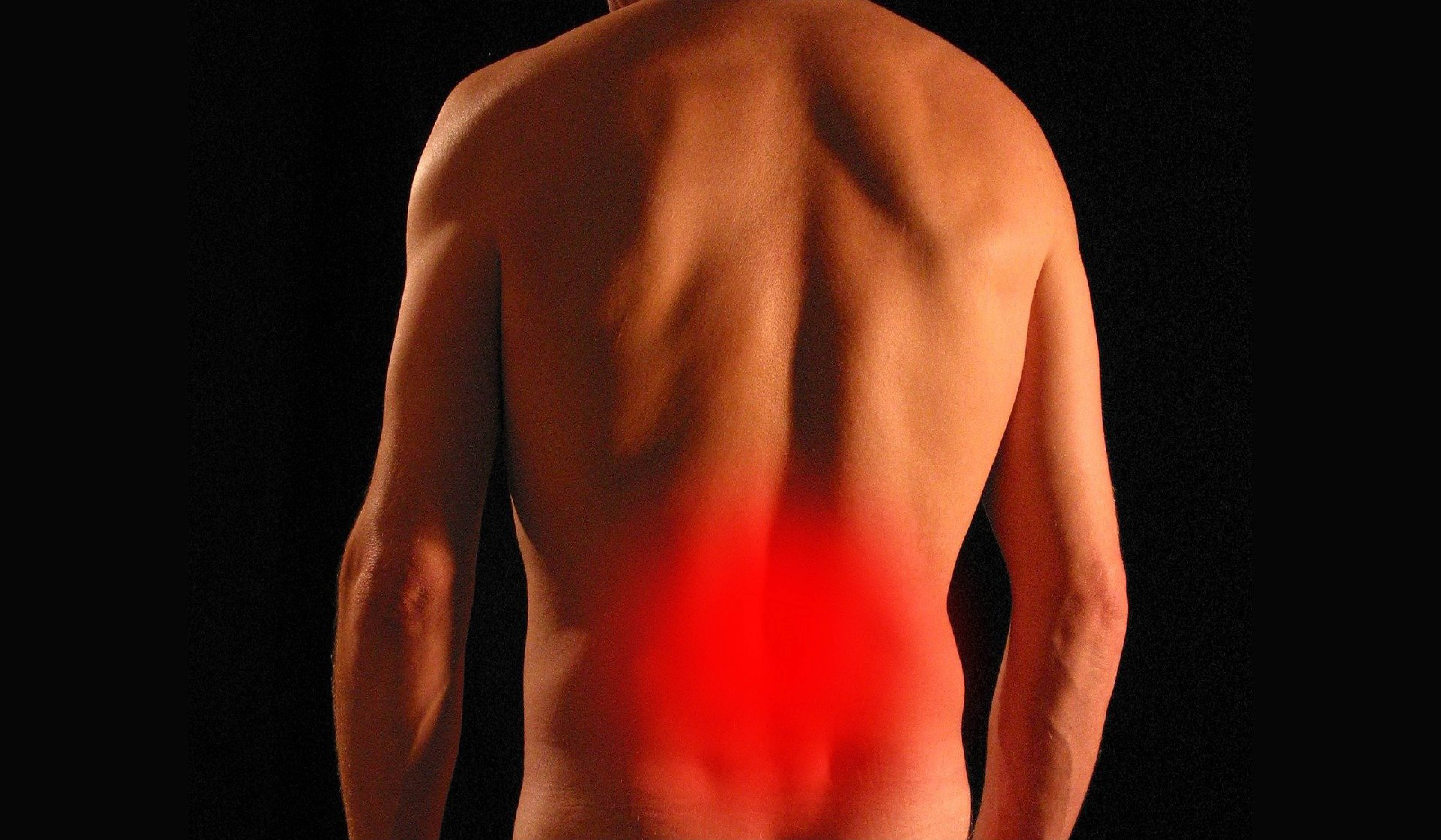
Our body has an abundance of healing pathways for self-repair.
Many of these self-repair mechanisms can be clinically manipulated for the creation of new therapies, making use of the body’s own self-healing potential.
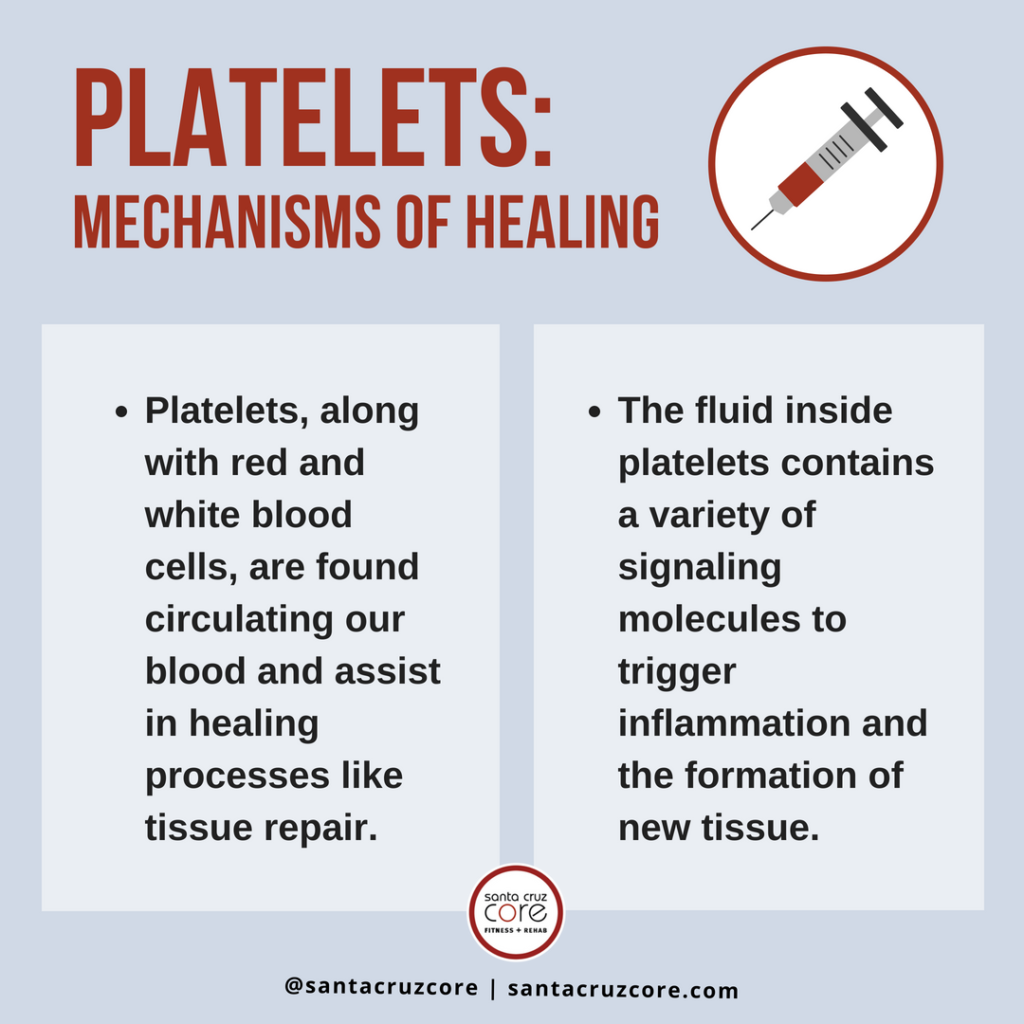
Platelets, normally found circulating in our blood, bear many of these self-healing mechanisms to clinically manipulate and trigger tissue repair.
Prolotherapy, for example, uses platelet-rich plasma solutions. Prolotherapy is an injection-based regenerative treatment used to treat musculoskeletal pain and related injuries. Rejuvenation of dermal tissue also uses platelet-rich plasma, as seen in the popular “vampire” facials.
The following represents a more in-depth narration on platelets, their healing mechanisms, and their therapeutic applications.
What Are Platelets?
Platelets normally reside circulating in our blood, along with red blood cells (RBCs), white blood cells (WBCs), and a variety of other cell types and molecules. All these cells are floating around in a fluid solution known as the blood plasma. In the blood plasma, we find a variety of other molecules including proteins, glycoproteins, carbohydrates, and fatty acids. Many of these molecules can be used to nourish cells or to act as chemical signals.
Together, all of these components make up what we refer to as blood.
This composition of blood allows for many its self-repair properties, assisting in the healing of vascular injuries, that is, an injured blood vessel. When a vascular injury is present, platelets adhere (or attach) to the site of injury where they come in contact with and are activated by collagen and other components of the vascular lining.
Once active, platelets themselves begin secreting signaling molecules that recruit more platelets and other coagulatory/inflammatory compounds. Eventually, platelets and these other compounds form a complex (or a plaque) to patch the injury and prevent further blood loss. This process of vascular repair is known as hemostasis (1).
Why is Platelets Hemostasis Significant?
During the process of hemostasis, platelets help recruit other components of healing through the release of signaling molecules like cytokines (which signal for inflammation). Eventually, this leads to the repair of the vascular injury and everything returns to normal. But is this the extent of platelet healing potential? Far from it.
Platelets are pockets of cytoplasm (the fluid inside of a cell) that break-off from much larger cells known as megakaryocytes. The fluid inside platelets contains a variety of signaling molecules that will trigger inflammation and the formation of new tissue when activated.
Signaling molecules found in platelets include cytokines and growth factors. They induce a healing cascade not limited to the vascular endothelium (the inside of blood vessels).
Thanks to this property, platelets can induce the repair of other tissue types including connective (soft) and dermal tissues. To better understand why these signaling molecules induce healing we must further discuss their properties.
Cytokines, Growth Factors, & Tissue Repair:
In molecular biology, the terms cytokines and growth factors get thrown around a lot. Many times, it ignores the lack of common knowledge for them. What exactly are they? Well, both of these molecule types represent a type of chemical signal. One single cell produces and releases these molecules. They then go on to stimulate and affect another. When chemical signals stimulate cell surface receptors, it alters their biochemical activities and growth.
Growth factors:
These are biochemical cues (or signals) that interact with cell surface receptors and exert an influence on that cell or tissue. Influences include cellular activation (to release more signals), maturation, and proliferation (making more cells) to name a few.
Cytokines:
Cytokines are a type of growth factor more specific to immune cells. These can stimulate their activation, maturation, and proliferation. For this reason, cytokines are most known for their roles in triggering inflammation, an immune response.
When exposed to an injured tissue, platelets’ own surface receptors activate and release more signaling molecules. These will then cascade down to more signaling reactions that will recruit and mature other cells necessary for tissue repair.
Specific Factors:
On December 2011, an issue of the Journal of Prolotherapy released an in-depth review of the mechanisms of PRP prolotherapy. The purpose of this publication was to explain the scientific evidence, methods, and applications of prolotherapy for the treatment of musculoskeletal pain (2).
When PRP is injected in soft tissue to induce an inflammatory healing cascade, several growth factors come into play. Growth factors involved in healing include (but are not limited to):
Platelet-derived growth factor (PDGF):
There are variations of this growth factor and it has a proliferative effect on many mesenchymal (adult) stem cell types which can eventually become specialized cells like osteoblasts (bone cells) and adipocytes (fat cells) (3).
Stromal cell-derived growth factor 1 alpha (SDF-1):
This growth factor supports adhesion and migration of mesenchymal stem/stromal cells (2). Stem cells are cells with less a determined function. They can take over the functions of other specialized cell types thus replacing them.
Epidermal growth factor (EGF):
This growth factor induces proliferation of mesodermal and ectodermal cell types, stem cells found in the middle and outermost layer of a developing embryo (3). These dermal layers eventually become specialized tissues of many cell types including those for the formation of muscle, connective tissue, skin, and nerves. It has a determining effect on stem cells to become specialized cells of the desired tissue.
Fibroblast growth factor (FGF):
There are many members of this family of growth factors, including 6 FGF families and even more FGF subfamilies (3). FGF plays a significant role in the development of the skeletal and nervous systems in mammals. Current tissue regenerative applications for FGF includes dermal, vascular, soft, adipose, and nervous tissues (4).
Insulin-like growth factor (IGF):
As the name entails, this growth factor has properties similar to insulin. It can even sometimes bind to insulin receptors, although at a much lower affinity (3). There are multiple IGF’s including IGF-1 and IGF-2. IGF-1 is a cellular response to the growth hormone (GH) and helps activate cascading pathways. IGF-2 is heavily expressed during fetal development and in a concept known as metabolic programming which helps determine organ function in adulthood. IGF-2 also assists in the activation of immune components necessary for the repair and healing of tissue.
When we take into account the effect of these growth factors individually and collectively, we can begin to understand how PRP helps repairs incompetent structures (such as injured soft tissue). Additionally, the inflammation healing cascade recruits a variety of healing mechanisms of its own.
Santa Cruz CORE offers prolotherapy and facial rejuvenation which utilizes platelet-rich plasma. Schedule a consultation today and see the real and true healing power of platelets!
References:
Parham, Peter, and Charles Janeway. The Immune System. Third ed., Garland Science, 2009.
Hauser, Ross A, et al. “Journal of Prolotherapy International Medical Editorial Board Consensus Statement on the Use of Prolotherapy for Musculoskeletal Pain.” Journal of Prolotherapy, 14 Aug. 2017, journalofprolotherapy.com/journal-of-prolotherapy-international-medical-editorial-board-consensus-statement-on-the-use-of-prolotherapy-for-musculoskeletal-pain/.
King, Michael W. “Growth Factors and Other Cellular Regulators.” TheMedicalBiochemistryPage, themedicalbiochemistrypage.org/growth-factors.php.
Yun, Ye-Rang, et al. “Fibroblast Growth Factors: Biology, Function, and Application for Tissue Regeneration.” National Center for Biotechnology Information, Journal of Tissue Engineering, 7 Nov. 2010, www.ncbi.nlm.nih.gov/pmc/articles/PMC3042641/.
